Before a single animal appeared in a museum case, scientists at the American Museum of Natural History set off on long expeditions to study wildlife in the field. They traveled to remote regions, enduring harsh weather and rough terrain. Their job was to observe animals in their natural habitats and return with skins, bones, plants, and detailed photographs. Every note and image they recorded would later serve as a blueprint for building accurate exhibits back in New York.
Specimens collected on these trips were carefully packed and shipped back to the museum. Once they arrived, the taxidermy department took over. Taxidermists in the 1930s and 1940s were both skilled scientists and artists. Their first task was cleaning and chemically treating the animal skins to stop decay and ensure they would last for decades.
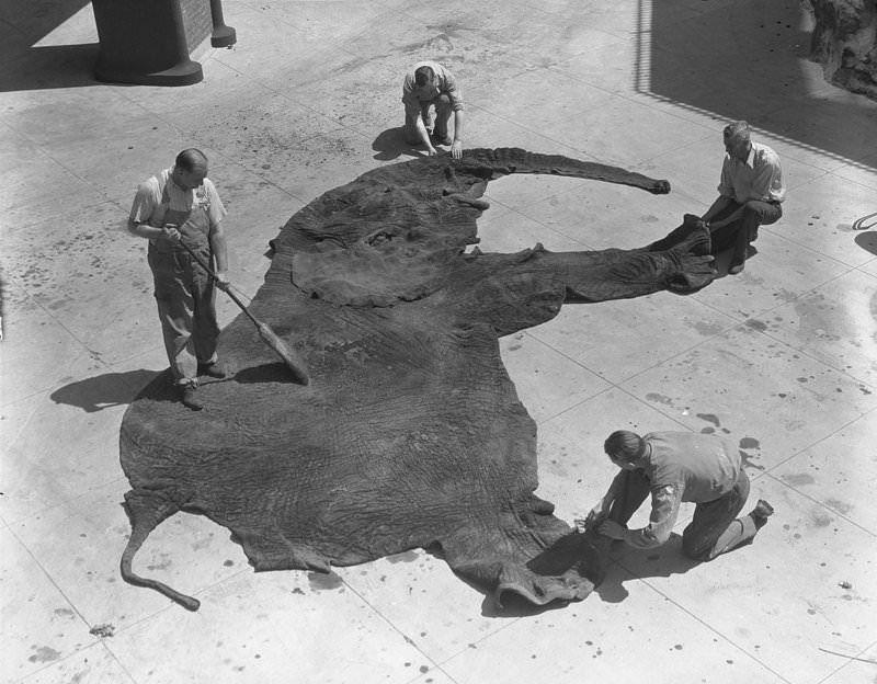
After preserving the skin, taxidermists built an internal body form called a manikin. Early manikins were constructed from wood, wire, and plaster, shaped to the exact dimensions of the original animal. The preserved skin was then stretched over the manikin and sewn into place with strong thread, leaving no wrinkles or gaps.
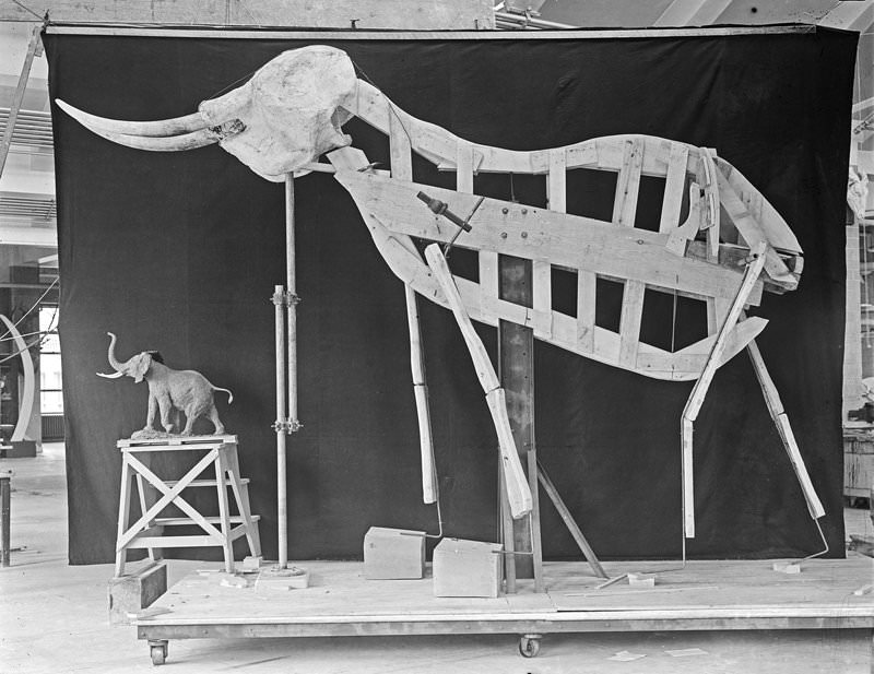
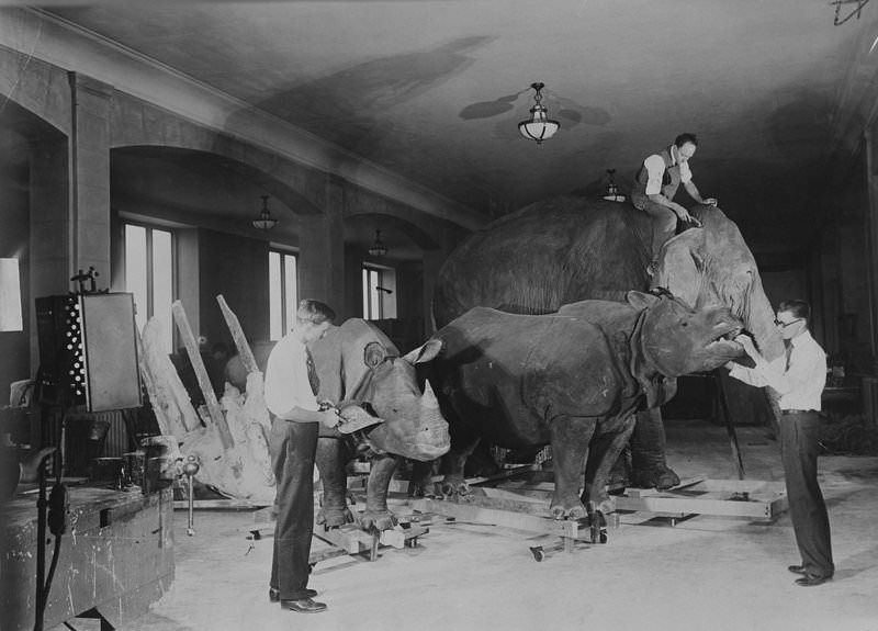
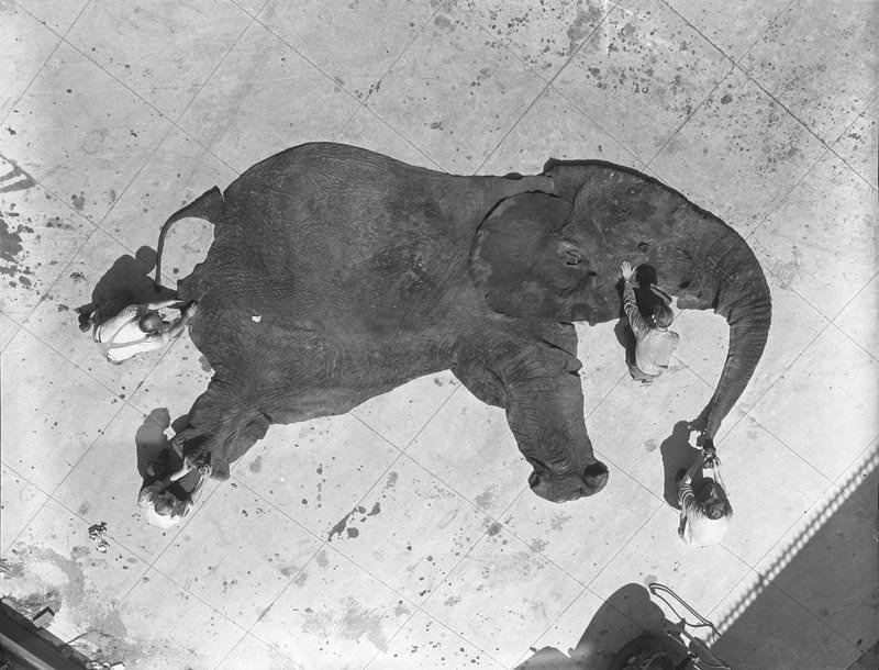
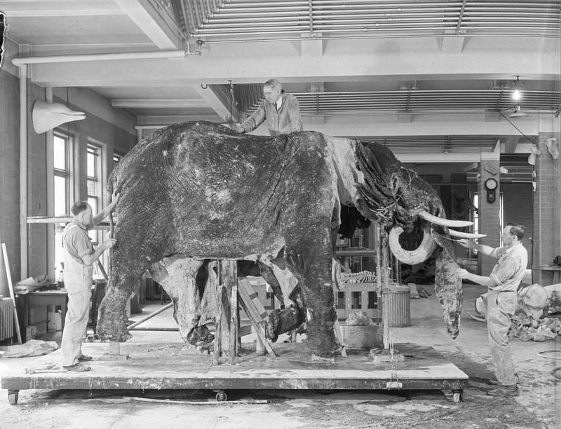
Large animals like elephants required an enormous amount of labor. Teams of staff worked together to lift, position, and secure massive skins onto their frameworks. Every pose was chosen to reflect a natural moment from the animal’s life in the wild.
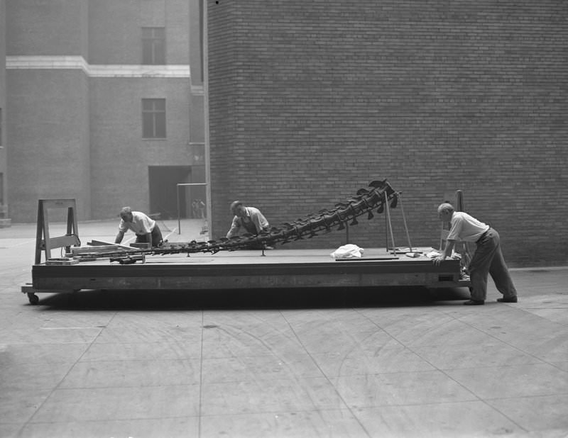
The work extended beyond mammals. Staff assembled full dinosaur skeletons from fossil specimens, a process that demanded equal precision. Moving large bones without causing damage required careful planning and steady hands.
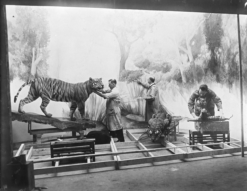
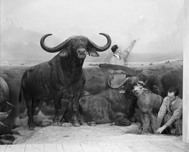
Group dioramas, like the African Buffalo Group, went far beyond mounting a single animal. Staff arranged multiple specimens together in scenes that told a story about animal behavior and habitat. Background paintings were added to complete the illusion of a real landscape.
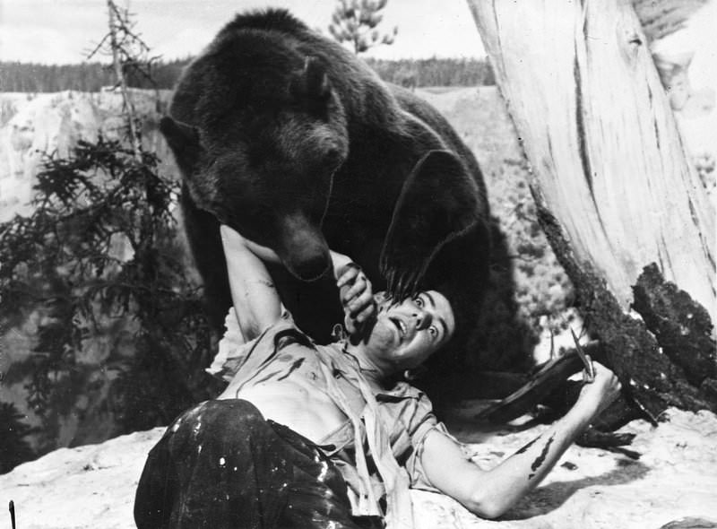
Some exhibits called for dramatic, action-filled poses. Grizzly bear groups, for example, were staged to show predatory behavior mid-motion. Capturing that energy in a static mount required taxidermists to study animal anatomy and movement in great detail.
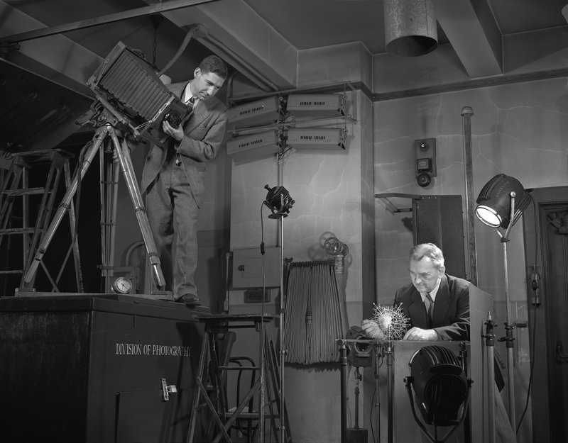
Not all exhibit work involved large animals. Specialists like glassblower Herman O. Mueller and photographer Charles H. Coles worked at a microscopic scale, recreating and documenting tiny organisms such as radiolarians for display.
The final stage brought all the elements together. In halls like the Asian Hall, staff painted large background murals and positioned mounted animals in front of them. Glass eyes, paint, and sculpted details gave each creature a lifelike presence. The result was an environment visitors could study as though standing in the wild themselves.